
Wounds that follow a burn can stay fragile for years. The scar tissue is thin, poorly supplied with blood, and easily breaks down with everyday movement, especially over a joint. When such a wound stops healing, the surgical goal is simple to state and harder to achieve: build a clean, healthy bed and then cover it with durable skin.
This case study follows one such patient through a single operation in which a collagen wound matrix (Rapiderm™) and a split-thickness skin graft were used together. It explains what was done at each step and why, and it is illustrated with six photographs taken during the procedure.
Patient Profile & Case Summary
| Presentation | Long-standing post-burn wound involving the forearm, extending towards the elbow |
| Patient | 66-year-old female |
| Setting | Fracture Clinic, Varanasi, Uttar Pradesh, India |
| Treating surgeon | Dr. Sudhir Singh (Plastic Surgeon) |
| Procedure | Surgical debridement, Rapiderm™ (Type I porcine atelocollagen wound matrix) application, split-thickness skin grafting (STSG) and a negative-pressure wound therapy (NPWT) dressing |
| Date of surgery | 30 September 2026 |
| Rapiderm applied | 30 September 2026 |
| Documentation | Intra-operative photographs, 30 September 2026 |
History and Presentation
A 66-year-old woman presented to the Fracture Clinic, Varanasi, under the care of Dr. Sudhir Singh (Plastic Surgeon), with a long-standing post-burn wound over the forearm that extended towards the elbow. The wound had the typical features of a chronic defect: irregular margins, patchy and unhealthy tissue quality, and no meaningful progress towards re-epithelialisation despite conservative dressing care.
Chronic wounds of this kind tend to stall because the underlying tissue has a poor blood supply and carries a layer of fibrotic or non-viable material that blocks new skin from growing across. Dressings alone cannot fix that. Given the size of the defect and how long it had been present, the surgical plan was debridement followed by wound-bed preparation with a collagen matrix and coverage with a split-thickness skin graft.
Understanding the Treatment Approach
The operation combined three ideas, each solving a different problem.
- Debridement removes dead, scarred and fibrotic tissue until a bleeding, healthy surface is exposed. Without this step nothing placed on the wound can take.
- Rapiderm™, a Type I porcine atelocollagen wound matrix, is laid over the prepared bed. A collagen matrix acts as a temporary scaffold: it protects the raw surface and gives the patient’s own cells and blood vessels a framework to grow into, which helps in irregular or marginally vascularised beds.
- A split-thickness skin graft (STSG) takes the top layers of skin (the epidermis and part of the dermis) from a donor site and places them over the wound. It gives fast, durable coverage and lets the donor site heal on its own.
Used together, the matrix helps the bed accept the graft and the graft closes the wound. Both were carried out on the same day.
Operative Management (30 September 2026)
Step 1: Preparation and debridement
The limb was prepped and draped in standard sterile fashion. Thorough debridement then removed the non-viable and fibrotic tissue until a clean, vascularised surface remained, which is the only kind of bed a graft can reliably take on. Fig. 1 shows the result: a raw, red, freshly bleeding surface spanning the forearm, with deeper soft tissue visible and sutures at the lower margin of the wound. The spots of fresh bleeding are a reassuring sign, because they show the bed has a living blood supply.
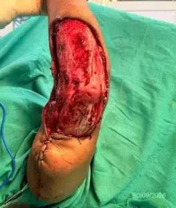
Fig. 1: The debrided recipient bed on the day of surgery (30/09/2026). The surface is clean, red and bleeding; no unhealthy or fibrotic tissue remains.
Step 2: Application of Rapiderm™
With the bed prepared, the Rapiderm™ sheet was placed directly onto the wound. In Fig. 2 the white matrix is being laid over the defect and smoothed down, with its edges still lifting at the margins. Fig. 3 shows it a little later: the matrix has absorbed moisture from the bed and moulded to the contours of the wound, and the pink-red tissue beneath shows through the thin, closely adherent layer. A matrix that sits flat and in full contact with the bed, without trapped fluid or air pockets, is what allows the scaffold to do its work.
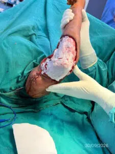
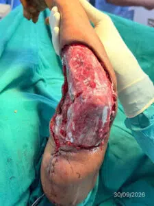
| Fig. 2: Rapiderm™ matrix being laid over the debrided forearm wound. | Fig. 3: The matrix moulded to the wound bed, with the tissue beneath showing through. |
Step 3: Skin Graft inset and fixation
A split-thickness skin graft was then harvested and inset over the defect. Fig. 4 shows the graft in place at the close of the procedure. Fine perforations are visible across its surface; these let blood and serum escape rather than collect under the graft and lift it off the bed. The graft was fixed all the way around its edge with skin staples. Keeping the graft in firm, uninterrupted contact with the bed is the single most important factor in whether it survives, so secure fixation is never rushed.
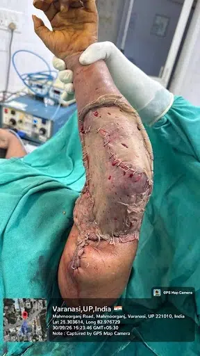
Fig. 4: Split-thickness skin graft inset over the forearm and secured circumferentially with skin staples (30/09/2026, 15:23).
Step 4: Final check of the fixed graft
With fixation complete, the limb was raised for final documentation (Fig. 5), taken seconds after the previous photograph. The graft is still uncovered here, so the staple line and the overall fit of the graft over the defect can be seen clearly before the dressing goes on.
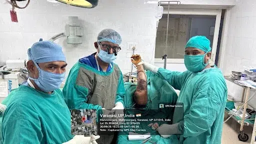
Fig. 5: The operating team with the grafted limb raised before dressing (30/09/2026, 15:23).
Step 5: Negative-pressure dressing
The graft was then covered with a negative-pressure wound therapy (NPWT) dressing, shown in Fig. 6 with the elbow flexed. A layer of foam sits over the graft and is sealed under a transparent adhesive film. A small port in the film connects through tubing to a collection canister, so that gentle suction can be applied. Over a graft, this does several useful things at once: it holds the graft evenly against the bed, draws away blood and serum before they can collect underneath, and limits shear, which matters a great deal over a joint that bends. The dressing was applied over non-adherent paraffin gauze against the graft, with a protective outer layer, to reduce shear and contamination during the first days.
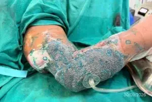
Fig. 6: Negative-pressure wound therapy dressing over the grafted forearm and elbow: foam under transparent film, with the suction port and tubing leading to the canister (30/09/2026).
What Makes a Graft Take
Whatever the technique, a graft survives only when a few conditions are met together:
- A clean, vascularised bed: all non-viable tissue is removed so the graft has living tissue to bond with.
- Good haemostasis: a blood or fluid collection under the graft pushes it away from the bed and can cause it to fail.
- Secure fixation: staples, sutures or dressings keep the graft from sliding, since shear tears away the fragile new connections. A negative-pressure dressing, as used here, adds even pressure across the whole graft.
- Early immobilisation and protection: especially important over a joint such as the elbow, where movement can disrupt the graft.
- Infection control: bacteria on the bed compete with the graft and are a common cause of loss.
Post-Operative Plan and Follow-Up
Photographs for this case were taken on the day of surgery, so no healing findings are reported here yet. In a case like this, the usual course is as follows.
- The first dressing change is typically done around post-operative day 5 to 7. The negative-pressure dressing is usually left in place until then, and when it is removed, the graft is inspected for adherence, fluid collections and signs of infection.
- Early graft take shows as a pink, well-perfused surface as new blood vessels grow into the graft from the bed (a process called inosculation, usually visible between days 4 and 7).
- Darker patches at the edges can appear and often settle with continued observation, but they are monitored.
- Staples are generally left until the graft is clearly stable, commonly around 10 to 14 days.
- Longer-term care includes scar management and early supervised movement once the graft has matured, to help prevent contracture across the elbow.
Discussion
Post-burn wounds over the forearm and elbow are challenging for a reason beyond poor healing. This region bends constantly, so grafts here face more movement and shear than almost anywhere else, and a raw area left to close slowly can tighten into a contracture that limits function. That makes both the quality of the bed and the stability of the graft especially important.
The strategy in this case was to address each risk directly: thorough debridement for a healthy base, a collagen matrix to support the bed, and a securely stapled graft for definitive cover, and a negative-pressure dressing to hold the graft steady. The photographs capture that sequence clearly, from a raw debrided bed to a fixed, perforated graft under a sealed NPWT dressing.
This is a single case, documented on the day of surgery, and it cannot by itself show how well the approach works. The graft outcome, the final appearance and the functional range of movement at the elbow will be the true measures of success and will be reported once follow-up is available.
Conclusion
This case follows a logical, stepwise pathway for a chronic post-burn forearm wound in a 66-year-old woman treated by Dr. Sudhir Singh (Plastic Surgeon) at Fracture Clinic, Varanasi: debridement to a clean bed, Rapiderm™ to support it, a split-thickness skin graft fixed securely with staples, and a negative-pressure dressing to protect it. The key lessons are that preparation of the bed matters as much as the graft itself, that fixation protects the graft during its most vulnerable days, and that wounds over joints need especially careful follow-up.